First Metatarsophalangeal Joint Arthritis: Comprehensive Review of Conservative and Surgical Treatment Options
Introduction
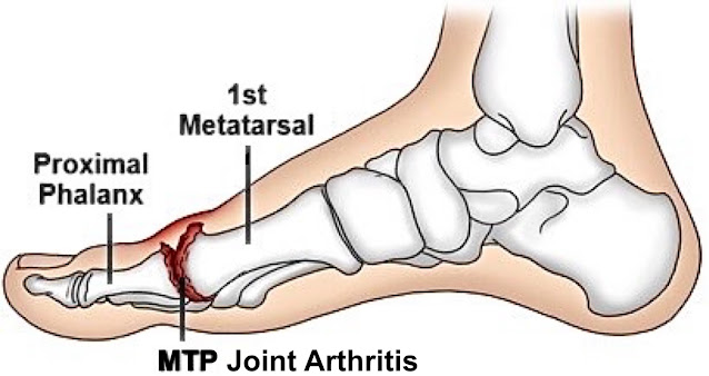
Arthritis of the first metatarsophalangeal (MTP) joint, commonly referred to as hallux rigidus when stiffness predominates, is a progressive degenerative condition affecting the articulation between the first metatarsal head and the proximal phalanx of the hallux. It is the most common arthritic condition of the foot and represents the second most common pathology of the first MTP joint after hallux valgus. The condition can significantly impair gait, athletic performance, occupational function, and overall quality of life.
First MTP joint arthritis is characterized by progressive degeneration of the articular cartilage, osteophyte formation, joint space narrowing, pain, and loss of motion. Dorsiflexion is typically affected first, resulting in altered gait mechanics and compensatory loading patterns throughout the foot and lower extremity.
The etiology of first MTP arthritis is multifactorial and may include repetitive microtrauma, prior injury, anatomical predisposition, inflammatory arthropathy, osteochondral lesions, biomechanical abnormalities, and genetic factors. The condition affects both active and sedentary populations and may present across a broad age range.
This article provides a comprehensive review of first MTP joint arthritis, including clinical presentation, diagnosis, conservative management strategies, and the full spectrum of surgical treatment options.
Anatomy and Biomechanics
The first MTP joint is a synovial hinge joint formed by the articulation of the first metatarsal head and the base of the proximal phalanx. The sesamoid apparatus beneath the first metatarsal head plays a critical role in load distribution and mechanical efficiency during gait.
Normal first MTP joint motion includes approximately:
65–75 degrees of dorsiflexion
15–30 degrees of plantarflexion
During the propulsive phase of gait, the first MTP joint experiences substantial forces, often exceeding body weight multiple times. Progressive cartilage degeneration and dorsal osteophyte formation restrict dorsiflexion, leading to compensatory gait changes such as lateral weight transfer, early heel rise, and increased forefoot loading.
Etiology and Risk Factors
Primary Osteoarthritis
Primary degenerative arthritis develops gradually without a clearly identifiable inciting event and may be related to repetitive stress and genetic predisposition.
Post-Traumatic Arthritis
Prior injury is a common cause and may include:
Turf toe injuries
Intra-articular fractures
Sesamoid injuries
Repetitive microtrauma
Osteochondral defects
Anatomical and Biomechanical Factors
Several structural abnormalities may predispose patients to first MTP degeneration:
Long first metatarsal
Metatarsus primus elevatus
Flat or chevron-shaped joint morphology
Hypermobile first ray
Hallux valgus interphalangeus
Pes planus or cavus deformity
Inflammatory Arthropathy
Inflammatory conditions that may affect the first MTP joint include:
Rheumatoid arthritis
Psoriatic arthritis
Gout
Seronegative spondyloarthropathies
Clinical Presentation
Patients commonly present with:
Pain localized to the dorsal or dorsomedial first MTP joint
Pain during push-off and walking
Progressive stiffness
Difficulty with stairs, running, or squatting
Swelling
Shoe irritation from dorsal osteophytes
Reduced athletic tolerance
As disease progresses, patients may alter gait mechanics to avoid painful dorsiflexion.
Physical Examination
Key examination findings include:
Reduced dorsiflexion
Dorsal osteophyte prominence
Pain at terminal dorsiflexion
Crepitus
Joint line tenderness
Swelling and synovitis
Altered gait mechanics
Transfer metatarsalgia
Advanced disease may demonstrate near-complete loss of motion and pain throughout the arc of movement.
Conservative Treatment Options
Conservative treatment is generally recommended initially for most patients unless severe deformity or end-stage arthritis is present.
The goals of nonoperative treatment include:
Pain reduction
Motion preservation
Functional improvement
Reduction of mechanical stress
Delay or avoidance of surgery
Activity Modification
Activity modification is often a first-line intervention.
Patients may benefit from reducing activities that require repetitive first MTP dorsiflexion, including:
Running
Sprinting
Lunges
Stair climbing
High-impact sports
Deep squatting
Low-impact alternatives include:
Cycling
Swimming
Elliptical training
Rowing
Footwear Modifications
Proper footwear can substantially reduce symptoms.
Recommended shoe characteristics include:
Wide toe box
Stiff sole
Rocker-bottom sole
Increased toe spring
Cushioned forefoot
Avoiding flexible minimalist shoes and high heels may reduce painful joint motion.
Orthotics and Bracing
Orthotic management aims to reduce painful first MTP motion and redistribute plantar pressures.
Custom Orthotics
Custom orthoses may include:
Morton’s extension
Carbon fiber foot plates
Reverse Morton’s extension
Arch support
Forefoot posting
Carbon Fiber Plates
Rigid carbon fiber inserts reduce first MTP dorsiflexion during gait and can provide meaningful symptom relief.
Turf Toe Plates
Rigid plates inserted into shoes help limit painful motion during athletic activities.
Physical Therapy
Physical therapy may provide symptom improvement in selected patients.
Potential interventions include:
Joint mobilization
Stretching exercises
Gastrocnemius stretching
Intrinsic foot strengthening
Gait retraining
Manual therapy
Balance training
Medications
NSAIDs
Nonsteroidal anti-inflammatory drugs may reduce pain and inflammation.
Examples include:
Ibuprofen
Naproxen
Meloxicam
Celecoxib
Topical Anti-Inflammatories
Topical diclofenac may provide localized symptom relief with reduced systemic exposure.
Acetaminophen
Acetaminophen may assist with pain control in patients unable to tolerate NSAIDs.
Ice and Cryotherapy
Cryotherapy can help reduce inflammation and pain, particularly after activity.
Typical recommendations include:
Ice application for 15–20 minutes
Several times daily as needed
Injections
Corticosteroid Injections
Intra-articular corticosteroid injections may provide temporary symptom relief.
Benefits include:
Reduction in inflammation
Short-term pain improvement
Diagnostic value
Limitations include:
Temporary duration
Potential cartilage toxicity with repeated injections
Risk of flare reaction or infection
Platelet-Rich Plasma (PRP)
PRP injections are increasingly utilized, though evidence remains mixed. Potential benefits include reduction in inflammation and symptom improvement.
Biologic and Regenerative Therapies
Additional investigational treatments include:
Bone marrow aspirate concentrate (BMAC)
Stem cell therapies
Amniotic-derived products
Long-term evidence remains limited.
Surgical Treatment Options
Surgical intervention is considered when conservative management fails to adequately relieve symptoms or when significant structural degeneration is present.
Surgical decision-making depends on:
Severity of arthritis
Patient age
Activity level
Functional goals
Motion preservation desires
Bone quality
Presence of deformity
Surgical procedures range from joint-preserving techniques to definitive fusion.


Comments